Epiretinal membrane treatment

Epiretinal membrane treatment
What is an epiretinal membrane?
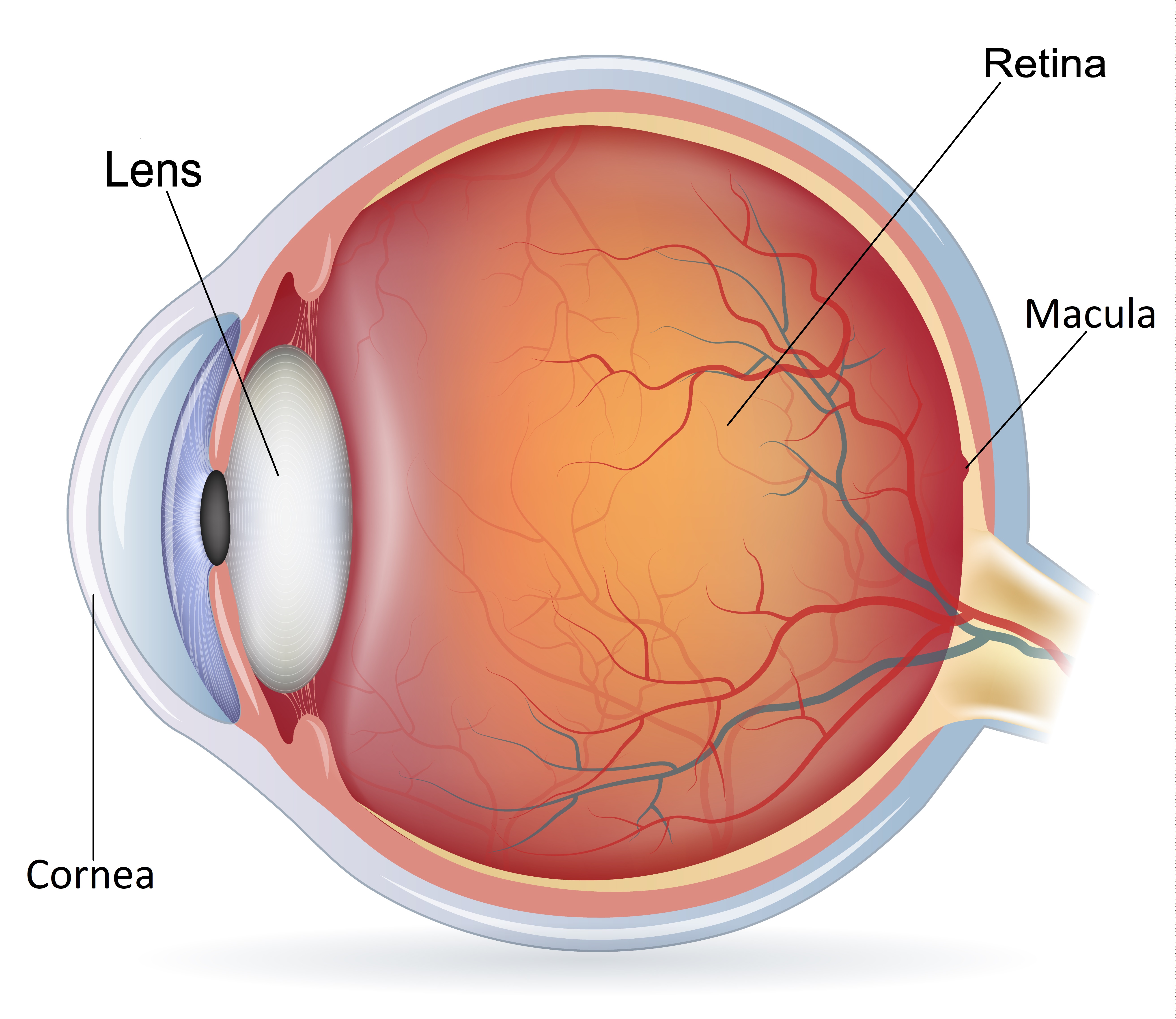
The eye is like a camera with a lens in the front and film at the back of the eye. The retina is the light sensitive film at the back of the eye. An epiretinal membrane is a condition where a thin layer of scar tissue forms on the surface of the retina. When this membrane grows over an area called macula (an important area responsible for our sharp central vision) on the retina, it causes wrinkling and crumples up the macula leading to distorted and/or blurred vision.
What causes epiretinal membrane?
In the majority of patients, epiretinal membrane develops due to the normal aging changes inside the eye. In some cases, it can be related to other conditions like diabetes, blockage of retinal blood vessels, inflammation or previous retinal surgery. Epiretinal membrane affects about 8% of people in later years. Epiretinal membrane is also called as Cellophane maculopathy or Macular pucker.
What are the symptoms
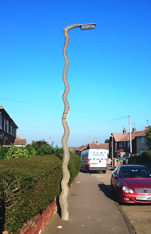
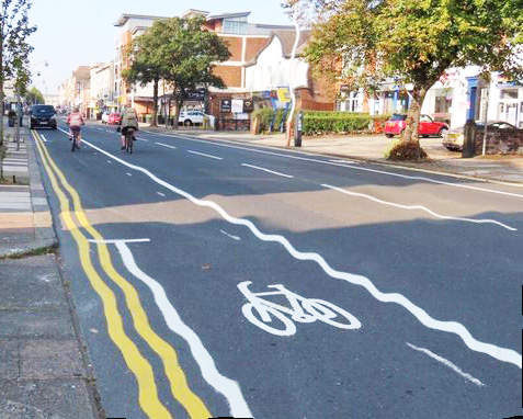
In many patients, epiretinal membrane is an incidental diagnosis made during routine eye examination and the vision may not be affected. However, epiretinal membrane can warp and wrinkle the retina which makes your vision to distort and blur, straight line may appear wavy and images may appear larger or smaller than real. These symptoms become obvious when good eye is covered.
Distorted vision - becomes pronounced when good eye is covered.
Epiretinal membrane removal surgery
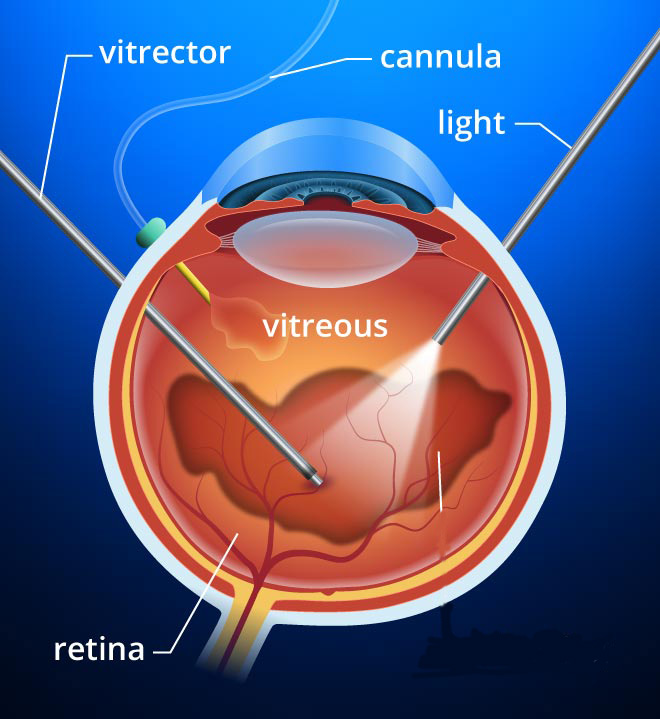
The epiretinal membrane is removed by an operation called a vitrectomy and epiretinal membrane peel. This involves a surgical procedure whereby the vitreous jelly is removed from inside the eye and the membrane is delicately peeled away from the central part of the retina. The operation can be performed under local or general anaesthesia as a day case procedure, which usually takes about an hour.
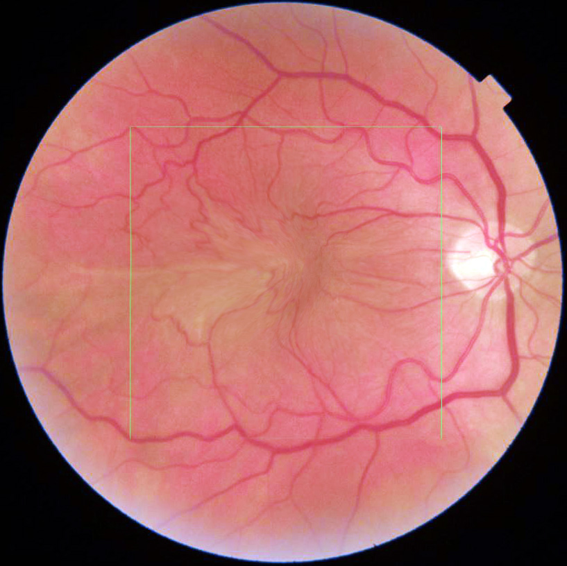
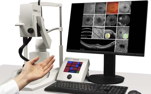
Though macular hole can be diagnosed by clinical examination, it is necessary to do OCT scan of retina, to get intricate details of macular hole to advice about the surgical outcome. Mr Viswanathan has access to the state-of-the-art retinal scanner to investigate the macular hole.
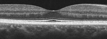
Cross-section of healthy Macula
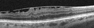
OCT scan of Epiretinal membrane
What to treat epiretinal membrane
In many patients, epiretinal membrane is an incidental diagnosis made during your routine eye examination and your vision may not be affected. These epiretinal membranes tend not to progress and do not affect your vision, so treatment may not be necessary. However, if they do get worse and if you have significant distortion and/or blurring of your vision which begins to affect yourquality of life then the only way to treat it is to remove the membrane surgically to reduce visual distortion.
Epiretinal membrane removal surgery – Sutureless Surgery
The epiretinal membrane is removed by aprocedure calledvitrectomy and epiretinal membrane peel. This involves a surgical procedure whereby the vitreous jelly is removed (Vitrectomy) from inside the eye to gain access to the membranefor it to be peeled away from the central part of the retina. The vitrectomy surgery is performed through three key-holes in the eye which does not require suturing.
In some cases, special gas bubble is left inside the eye which will dissolve on its own after few weeks. If gas is used, you will be asked to position your head in a certain way for some part of the day for up to 7 days and you must not fly or go high altitude until gas bubble disappears. The operation can be performed under local or general anaesthesia as a day case procedure, which usually takes about an hour. If there is a cataract along with epiretinal membrane, then cataract is also removed at the same time a single procedureduring epiretinal membrane surgery.
HOW SUCCESSFUL IS THE SURGERY
Epiretinal membrane peel surgery is successful in vast majority of patients, as 80 to 90 % of patients undergoing this surgery notice improved vision with a decrease in visual distortion. Much of the visual improvements occurs in the first few weeks and this can take up to 6 months
Where can I find further information?
Understanding epiretinal membrane surgery can be complicated. The information given above will not cover all the concerns you may have about this procedure. Further information can be found at the following websites:
https://www.rnib.org.uk/eye-health/eye-conditions/epiretinal-membrane
Information leaflet downloads
Epiretinal membrane leaflet Amsler Grid leaflet
Scientific Evidence
The information mentioned here is based on a variety of sources, including latest published research and the Britain & Eire Association of Vitreoretinal Surgeons.
Disclaimer
It is impossible to diagnose and treat patients without complete eye examination by an ophthalmologist. I hope the above information will be of help before and after a consultation which this information supplements and does not replace. This information must not be used as a substitute for professional medical care by a qualified doctor or other health care professional.
If you have any concerns about your condition or treatment, please ask your surgeon Mr Viswanathan. We are not responsible or liable, directly or indirectly, for ANY form of damages whatsoever resulting from the use (or misuse) of information contained in this page or found on web pages linked to from this page.